
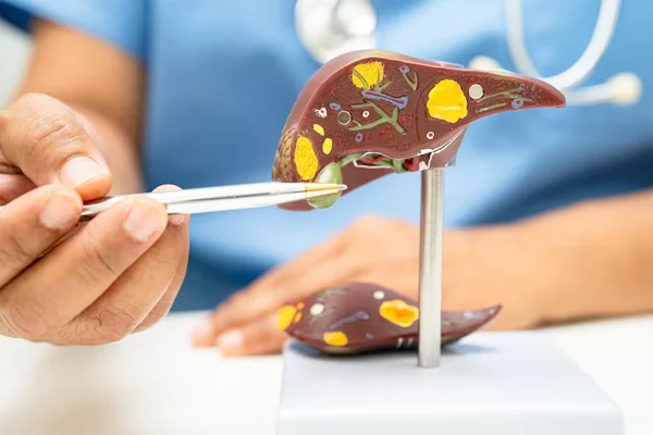
A liver biopsy involves collecting a small sample of liver tissue for laboratory examination. It helps assess liver inflammation, scarring, and other abnormalities when blood tests, imaging, or other investigations do not provide sufficient information.
Percutaneous liver biopsy involves inserting a biopsy needle through the skin, usually in the upper right side of the abdomen, to obtain a small tissue sample. Imaging guidance may be used to help select an appropriate insertion site. This approach is considered when the patient's clinical condition and blood clotting status make it suitable.
Transjugular liver biopsy involves passing a catheter through a vein in the neck and guiding it into the hepatic venous system to collect liver tissue. It may be preferred for selected patients with an increased bleeding risk, significant fluid accumulation in the abdomen, or other conditions that make the percutaneous approach less suitable.
The collected tissue is examined under a microscope to help diagnose or assess liver diseases. The choice of technique depends on the patient's medical condition, laboratory results, imaging findings, and individual procedural risks.